What is liver?
The liver is a vital organ in the human body, playing a central role in various bodily functions. The liver is located in the upper right side of the abdominal cavity, below the diaphragm, and is divided into four lobes. It receives blood from the hepatic artery and hepatic portal vein, and its waste products are excreted through the bile ducts into the intestines.
Anatomy of liver
The liver is a complex organ with a unique anatomy, consisting of:
1. Location: Upper right side of the abdominal cavity, below the diaphragm.
2. Shape: Irregular, triangular shape with four lobes (right, left, caudate, and quadrate).
3. Size: Approximately 3 pounds (1.4 kg) in adults.
4. Surface:
a. Diaphragmatic surface: Faces the diaphragm.
b. Visceral surface: Faces the abdominal cavity.
1. Lobes:
a. Right lobe: Largest lobe, divided into anterior and posterior segments.
b. Left lobe: Smaller lobe, divided into medial and lateral segments.
c. Caudate lobe: Small, tail-like lobe.
d. Quadrate lobe: Small, square-shaped lobe.
1. Blood supply:
a. Hepatic artery: Supplies oxygenated blood.
b. Hepatic portal vein: Supplies nutrient-rich blood from the digestive tract.
1. Bile production and drainage:
a. Hepatocytes: Liver cells producing bile.
b. Bile canaliculi: Small channels collecting bile.
c. Bile ducts: Larger ducts merging to form the common hepatic duct.
d. Common bile duct: Drains bile into the small intestine.
1. Lymphatic system: Drains lymph from the liver into the thoracic duct.
2. Nerve supply: Innervated by the vagus nerve and sympathetic nervous system.
Understanding the liver’s anatomy is crucial for diagnosing and treating liver diseases and disorders.
Functions of liver
The liver performs numerous vital functions, including:
1. Detoxification: Removes toxins, waste products, and excess substances from the blood.
2. Metabolism: Breaks down carbohydrates, proteins, and fats, converting them into energy.
3. Production of bile: Produces bile to aid in fat digestion and absorption.
4. Storage of nutrients: Stores vitamins, minerals, and glycogen (a complex carbohydrate).
5. Blood filtration: Filters the blood, removing old or damaged red blood cells.
6. Hormone regulation: Regulates hormone levels, including insulin and thyroid hormones.
7. Immune system support: Supports the immune system by removing pathogens and toxins.
8. Production of proteins: Produces essential proteins, such as clotting factors and lipoproteins.
9. Maintenance of blood sugar levels: Regulates blood sugar levels by storing or releasing glucose.
10. Processing of medications: Metabolizes and eliminates drugs and medications.
11. Production of cholesterol: Produces cholesterol, essential for hormone production and cell membrane structure.
12. Storage of iron: Stores iron, essential for healthy red blood cells.
13. Removal of bilirubin: Removes bilirubin, a byproduct of red blood cell breakdown.
14. Supports gut health: Produces bile, which helps maintain a healthy gut microbiome.
The liver’s diverse functions make it a vital organ, and liver dysfunction can lead to various diseases and disorders.
What is liver transplant radiology
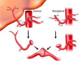
Liver transplant radiology refers to the use of medical imaging techniques to evaluate the liver and its blood vessels before, during, and after a liver transplant surgery. Radiologists use various imaging modalities to:
1. Evaluate liver anatomy: Assess the liver’s size, shape, and blood vessel structure.
2. Detect liver disease: Identify liver damage, cirrhosis, tumors, or other conditions.
3. Plan surgery: Guide surgeons during liver transplant procedures.
4. Monitor transplant function: Evaluate the transplanted liver’s blood flow, function, and potential complications.
When liver transplant radiology is necessary?
Liver transplant radiology is necessary in the following situations:
1. Pre-transplant evaluation: To assess the liver anatomy, detect any abnormalities, and evaluate liver function in potential recipients.
2. Donor liver evaluation: To assess the donor liver anatomy, detect any abnormalities, and evaluate liver function.
3. Transplant procedure guidance: To guide surgeons during the transplant procedure.
4. Post-transplant complications: To detect and evaluate complications such as:
– Vascular thrombosis or stenosis
– Bile duct obstruction or leakage
– Rejection or graft dysfunction
– Infection or abscess
5. Follow-up and monitoring: To regularly evaluate liver function and detect any potential complications.
6. Liver cancer treatment: To evaluate the extent of liver cancer and plan treatment.
7. Liver disease diagnosis: To diagnose and evaluate liver diseases such as cirrhosis, fatty liver disease, or liver fibrosis.
8. Bile duct disease: To evaluate and treat bile duct diseases such as primary sclerosing cholangitis.
9. Portal hypertension: To evaluate and treat portal hypertension and its complications.
10. Pediatric liver disease: To evaluate and treat liver diseases in children.
Liver transplant radiology is crucial for ensuring the success of liver transplant procedures and monitoring the health of the transplanted liver.
Procedure of liver transplant radiology
Here is a more detailed overview of the procedure of liver transplant radiology:
Pre-Transplant Evaluation
1. Ultrasound: Evaluate liver anatomy, detect any abnormalities, and assess liver function.
2. CT/MRI: Evaluate liver anatomy, detect tumors or disease, and assess blood flow.
3. Angiography: Visualize blood vessels and assess patency.
Donor Liver Evaluation
1. Ultrasound: Evaluate donor liver anatomy and detect any abnormalities.
2. CT/MRI: Evaluate donor liver anatomy and detect tumors or disease.
3. Angiography: Visualize donor liver blood vessels and assess patency.
Transplant Procedure
1. Intraoperative ultrasound: Guide surgeons during transplant procedure.
2. Angiography: Visualize blood vessels and guide surgeons during anastomosis.
Post-Transplant Evaluation
1. Ultrasound: Evaluate liver function, detect any complications, and assess blood flow.
2. CT/MRI: Evaluate liver function, detect any complications, and assess blood flow.
3. Angiography: Visualize blood vessels and assess patency.
Follow-Up
1. Ultrasound: Regularly evaluate liver function and detect any complications.
2. CT/MRI: Regularly evaluate liver function and detect any complications.
3. Angiography: Regularly evaluate blood vessels and assess patency.
Interventions
1. Biopsy: Evaluate liver function and detect any complications.
2. Angioplasty: Treat any vascular complications.
3. Stenting: Treat any vascular complications.
Note: The specific procedures and imaging modalities used may vary depending on the individual case and the transplant center’s protocols.
Side effects of liver transplant radiology
While generally safe, liver transplant radiology procedures can have some side effects, including:
1. Allergic reactions: To contrast agents used in imaging procedures.
2. Radiation exposure: From CT scans, which can increase cancer risk.
3. Bleeding or bruising: At biopsy or angiography sites.
4. Infection: At biopsy or angiography sites.
5. Nephrotoxicity: Contrast agents can harm kidney function.
6. Thrombosis or embolism: From angiography or biopsy procedures.
7. Hemorrhage: From biopsy or angiography procedures.
8. Liver damage: From biopsy procedures.
9. Gallbladder or bile duct damage: From angiography or biopsy procedures.
10. Anaphylaxis: A severe allergic reaction to contrast agents.
11. Hypersensitivity reactions: Mild to severe reactions to contrast agents.
12. Kidney failure: In patients with pre-existing kidney disease.
13. Contrast-induced nephropathy: Kidney damage from contrast agents.
14. Radiation-induced injuries: From prolonged radiation exposure.
15. Vascular injuries: From angiography or biopsy procedures.
It’s essential to discuss any concerns or potential risks with your radiologist or healthcare provider before undergoing liver transplant radiology procedures. They can help minimize risks and ensure your safety.
